
For patients who have lost all or most of their teeth, implant treatment can provide an alternative to conventional removable dentures. However, full arch implant rehabilitation involves more than simply placing implants. Accurate implant positioning, soft tissue information, jaw relationships, and prosthetic design all need to work together.
As digital dentistry continues to develop, a photogrammetry 3D scanner can help address one of the most demanding parts of this workflow: accurately recording the position and orientation of multiple implants. For clinicians and dental laboratories managing complex full arch cases, integrating photogrammetry with intraoral scanning can support a more streamlined approach to edentulous patient treatment.
Can Edentulous Patients Receive Dental Implants?
Being edentulous does not automatically prevent a patient from receiving dental implants. Depending on the patient’s bone volume, anatomical conditions, general oral health, and treatment objectives, implants may be used to support fixed or removable prostheses.
A complete arch implant case typically requires careful planning before treatment begins. CBCT data can provide information about the underlying bone and anatomical structures, while digital scans can capture the existing oral conditions and prosthetic surfaces. These datasets can then be combined during virtual treatment planning.
Research has demonstrated fully digital workflows for edentulous implant patients by integrating intraoral scanning with CBCT data, allowing clinicians to plan implant positions and design implant supported restorations digitally.
The challenge becomes particularly apparent when many implants are involved. Unlike natural teeth, edentulous arches provide fewer stable anatomical landmarks for conventional intraoral scanning. This can make the accurate registration of implant positions more difficult, especially across a complete arch.
Why Implant Position Accuracy Matters in Full Arch Cases
In a single implant restoration, small positioning discrepancies may be relatively easy to manage. In a full arch restoration supported by multiple implants, however, errors can accumulate across the arch.
The digital record needs to accurately communicate the three dimensional position and angulation of each implant. If the recorded implant positions do not correspond closely to the clinical situation, the resulting restoration may require additional adjustments or compromise the intended passive fit.
This is where dental photogrammetry can complement conventional intraoral scanning. Rather than relying only on the visual surface of the gingiva, photogrammetry can use coded scan bodies as reference points to calculate implant positions and orientations. The resulting implant information can then be combined with detailed intraoral surface data.
For edentulous patient treatment, this distinction is important because the implant location and the surrounding soft tissue both contribute to the final prosthetic design.
What Is a Photogrammetry 3D Scanner?
A photogrammetry 3D scanner captures the spatial relationship between multiple reference points and calculates their three dimensional positions. In implant dentistry, specially designed coded scan bodies can provide identifiable reference points for determining implant position and angulation.
Intraoral photogrammetry can therefore serve a different function from conventional intraoral scanning. The photogrammetry component focuses on implant position, while intraoral scanning captures teeth, gingiva, and other oral surface information.
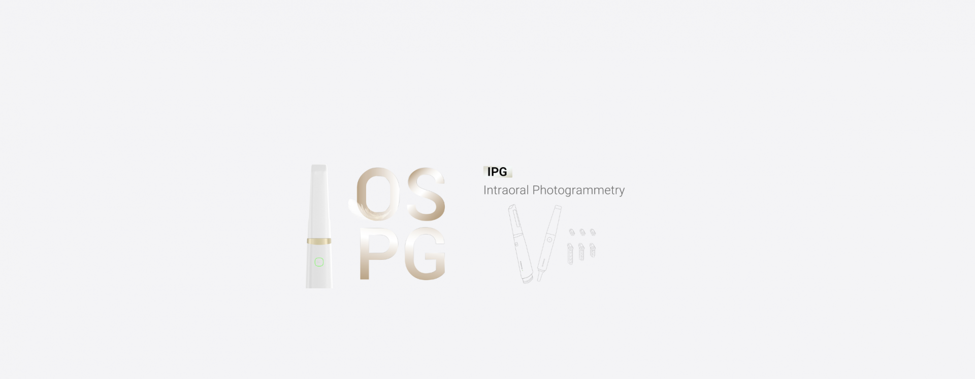
According to SHINING 3D DENTAL, its Intraoral Photogrammetry technology integrates photogrammetry with intraoral scanning. In edentulous and partially edentulous cases, the system can capture implant positions and angles together with intraoral and gingival data, providing digital information for implant supported restorations.
This combined approach can reduce the need to manage separate scanning systems and datasets during the digital workflow.
How Digital Scanning Supports Edentulous Patient Treatment
A typical digital workflow may begin with clinical examination and CBCT acquisition. Implant positions are planned according to anatomical conditions and prosthetic requirements. After implant placement, scan bodies can be used to record the implant positions.
The photogrammetry data can then be combined with intraoral scan data. These digital records provide the foundation for CAD design, communication with the dental laboratory, and fabrication of the final restoration.
For patients with an existing denture, the denture can also provide useful information about tooth arrangement, vertical dimension, and occlusion. Digital workflows have demonstrated that existing dentures and temporary prostheses can be incorporated into the digital process to help transfer important prosthetic information.
For clinicians, this means that digital treatment is not simply about replacing an impression. It is about connecting diagnosis, implant planning, prosthetic design, and manufacturing through a consistent data workflow.
SHINING 3D DENTAL and the Integrated IPG Approach
SHINING 3D DENTAL has developed an integrated approach through its Aoralscan Elite platform, which combines intraoral scanning and intraoral photogrammetry in one device. The company positions this workflow for implant cases including edentulous jaw rehabilitation and All on X treatment.
This integration can be particularly useful when a case requires both precise implant position information and detailed intraoral surface data. Instead of treating implant photogrammetry and conventional intraoral scanning as completely separate procedures, clinicians can acquire and merge the necessary datasets within a connected workflow.
Recent clinical research has also evaluated IPG based complete arch implant rehabilitation in fully edentulous patients, reflecting the growing clinical interest in photogrammetry based digital workflows.
Building a More Connected Full Arch Workflow
Successful edentulous implant treatment still depends on clinical diagnosis, implant planning, surgical execution, prosthetic design, and professional judgment. A scanner cannot replace these essential steps.
However, accurate digital data can help connect them. By combining CBCT, implant position records, intraoral scans, and prosthetic information, clinicians and dental laboratories can establish a more comprehensive digital representation of the patient.
For full arch implant cases, a photogrammetry 3D scanner offers a practical way to capture implant position information that can be difficult to obtain through conventional intraoral scanning alone. When integrated with a broader digital workflow, this technology can support more consistent data transfer, prosthetic planning, and edentulous patient treatment.
As digital dentistry continues to evolve, solutions such as SHINING 3D DENTAL provide an integrated pathway for bringing implant photogrammetry and intraoral scanning into the same clinical workflow.